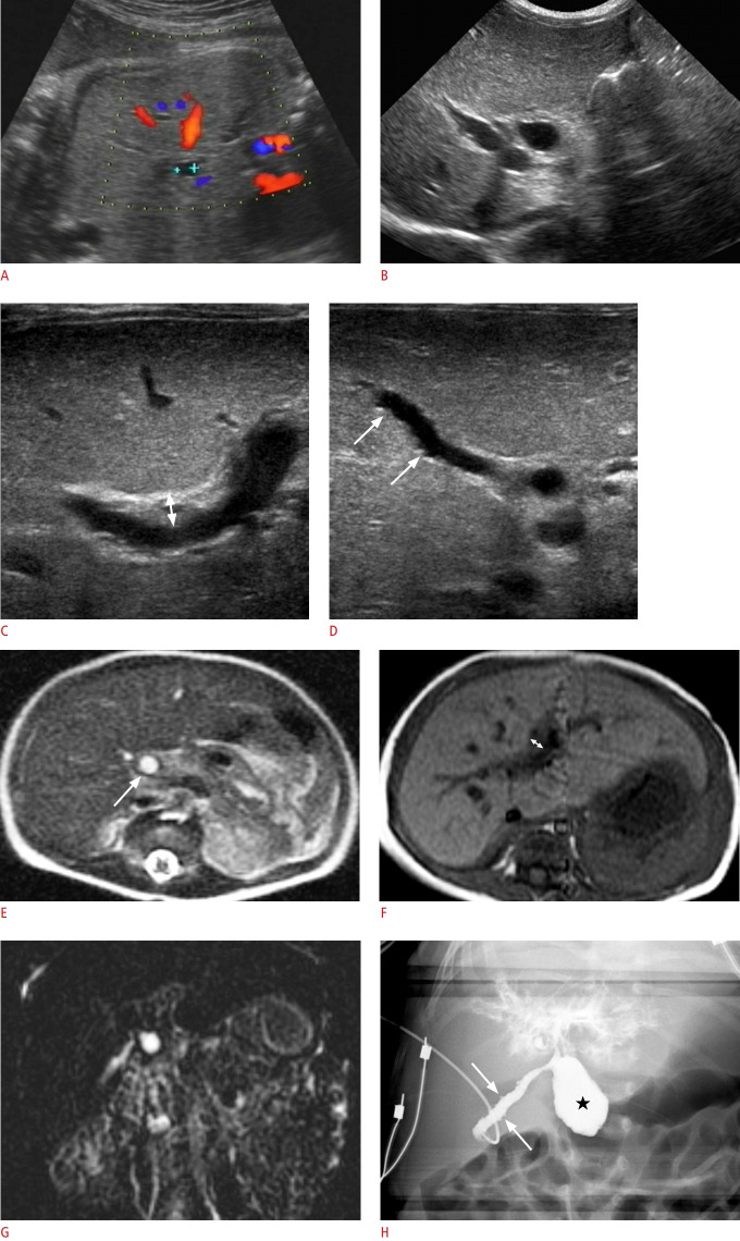
Fig. 1. A girl with cystic biliary atresia.
A. Prenatal ultrasonography (US) at a gestational age of 35 weeks shows a 0.6-cm cyst around the hepatic hilum. B-D. Abdominal US on the second day after birth shows the cyst (0.7 cm) at the hepatic hilum (B), increased periportal echo with triangular cord thickness (double arrow) of 4.8 mm (C), and mucosal irregularity (arrows) of the elongated gallbladder (GB) (D). E-G. Preoperative abdominal magnetic resonance imaging also demonstrates a hepatic hilar cyst (0.7 cm, arrow) on a T2-weighted axial image (E), periportal thickening with a triangular cord thickness (double arrow) of 4.0 mm on a T1-weighted axial image (F), and no visible distal common bile duct (CBD) on three-dimensional magnetic resonance cholangiopancreatography (G). H. Intraoperative cholangiography confirmed an elongated GB with mucosal irregularity (arrows) connected with the cystic lesion (star) in the proximal CBD, an invisible distal CBD, and contrast leakage at the hepatic hilar area without visible normal bile duct branches. She was confirmed to have cystic biliary atresia and underwent the Kasai operation.

