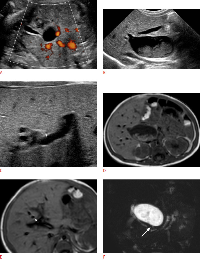
Fig. 2. A boy with choledochal cyst type Ia.
A. Prenatal ultrasonography (US) at a gestational age of 31 weeks shows a hepatic hilar cystic lesion (2.0 cm) and gallbladder (GB). B, C. Postnatal abdominal US also shows the cystic lesion (about 2.6 cm) at the hepatic hilar area with internal sludge, a grossly normal GB (B), and no remarkable periportal echo with a triangular cord thickness (double arrow) of 1.9 mm (C). D, E. T1-weighted axial magnetic resonance images demonstrates the same findings of the cyst (3.4 cm) with internal sludge (D) and periportal signal change with a triangular cord thickness (double arrow) of 3.9 mm (E). F. Magnetic resonance cholangiopancreatography shows patent distal common bile duct (arrow). The cystic lesion was confirmed as a choledochal cyst based on pathology findings.

