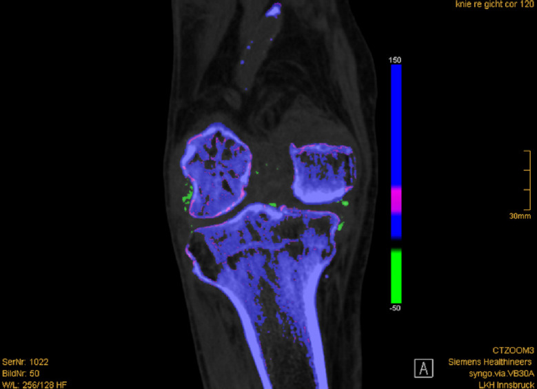
Fig. 4. Monosodium urate (MSU) deposit in a dual-energy computed tomography (DECT) of the knee.

Coronal DECT shows color-coded green MSU deposits in the right knee affecting the medial and lateral collateral ligaments, cruciate ligaments, and intracondylar fossa.
