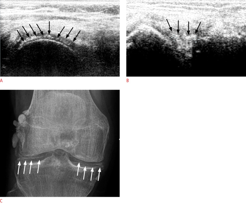
Fig. 5. A patient with calcium pyrophosphate dihydrate (CPPD) of the knee.
A. Axial ultrasonography (US) of the femoral condyle shows a thick linear deposit (arrows) within the cartilage layer, indicates high-grade CPPD. B. Parasagittal US of the lateral knee shows echogenic deposits (arrows) within the lateral meniscus. C. Corresponding X-ray of the same patient shows typical calcification (arrows) within the menisci and around the condyle.

