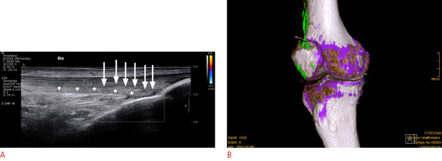
Fig. 7. Extra-articular monosodium urate (MSU) deposits in the quadriceps tendon.
A. Longitudinal ultrasonography of the distal quadriceps tendon is shown. No hypervascularity or tendinosis is seen, but there are diffuse discrete hyperechoic deposits at the posterior layer of the tendon (stars). There is no dorsal shadowing, but a discrete hypoechoic halo is present (arrows). The extent of the abnormality only became apparent retrospectively after performing dual-energy computed tomography (DECT). B. Sagittal three-dimensional rendered DECT demonstrates color-coded green MSU deposits along the quadriceps tendon and the prepatellar region. Furthermore, discrete MSU deposits (green) are visible in the infrapatellar tendon, medial collateral ligaments, and menisci.

