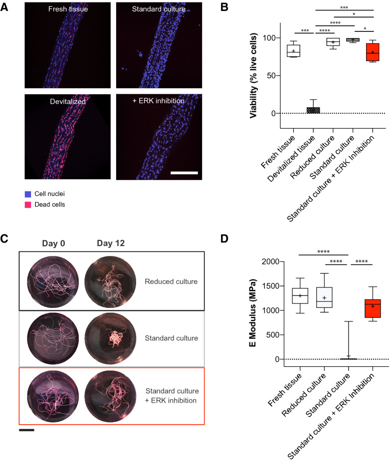
Figure 2.
Inhibition of ERK 1/2 activity prevents loss of tendon biomechanical properties in tendon explants cultured for 12 days. (A) Representative fluorescence microscopy images of tendon fascicles with nuclear staining of all cells (blue) and dead cells (EthD-1, violet). Scale bar 200 μm. (B) Quantification of cell viability in uncultured tendon explants (fresh tissue), methanol treated uncultured negative control tendons (devitalized tissue) and ex vivo cultured explants in reduced and standard culture treated without and with an ERK 1/2 inhibitor, N = 6. Devitalized tissue significantly differs from all other conditions. (C) Representative macroscopic images of tendon fascicles before treatment (uncultured tendon explants, day 0) and after 12 days of incubation in indicated culture conditions. Scale bar 10 mm. (D) Elastic moduli of isolated tendon fascicles (freshly isolated or cultured ex vivo in reduced conditions, standard conditions or treated with an ERK 1/2 inhibitor), N = 12 (N = 10 for standard culture, due to tissue rupture during the mounting procedure. Negative values of elastic modulus resulting from preliminary tissue failure were set to zero). The standard culture significantly differs from all other conditions. GraphPad Prism (version 8.4.3) was used to perform statistical analyses and generate the figures. Box plots (25th and 75th percentiles) with whiskers (5th and 95th percentiles), median (line) and mean (+). Bar plots represent mean values + SD. All statistical tests unless otherwise stated: Repeated measures ANOVA with Tukey's multiple comparisons test with *p < 0.05, ***p < 0.001, ****p < 0.0001.

