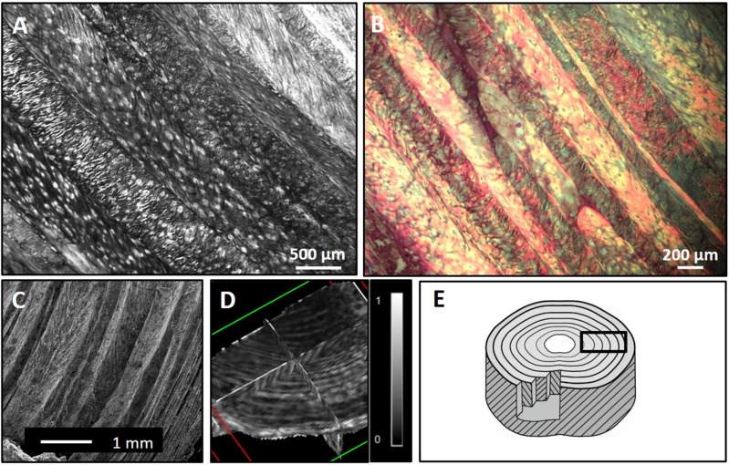
Figure 1.
A portion of the outer AF and its structure. Two-dimensional high-resolution histological images of the AF (axial section) using light microscopy (A, B): The different orientations of the fiber tracts in each lamella are clearly seen in (A), whereas (B) (Masson Trichrome stain) presents the complex structure of the AF. The 2D scanning microscope image yielded a consistent arrangement (C). DTI generated 3D FA data where distinct concentric lamellae rings are seen (as in the histological sections), since they are associated with anisotropic water diffusion (D). The scheme (E) shows lamellae layers with alternating orientation fibers surrounding the nucleus pulposus (center).

