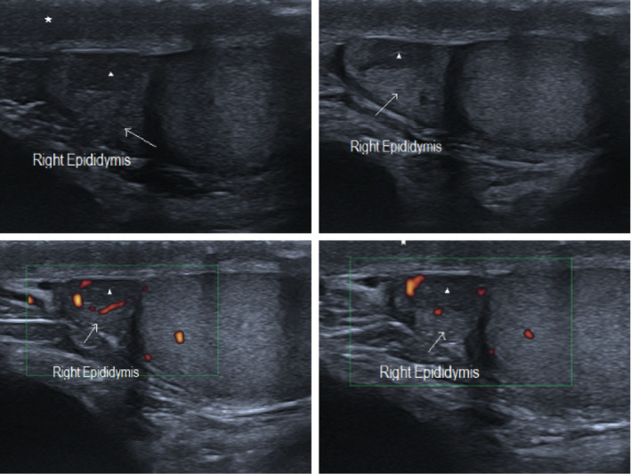
FIGURE 1.

SARS‐COV 2 subclinical epididymitis pattern #1: Epididymal head enlargement (arrow) with inhomogeneous echogenicity and increased colour Doppler blood flow. Marked hypoechogenic small areas with peripherical Doppler vascularization indicating micro‐abscess (arrowhead). Reactional oedema and thickness of the scrotum wall as well (asterisk)
