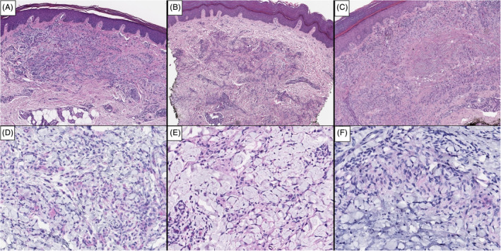
FIGURE 1.

Immunohistochemical staining of granuloma annulare in biopsies from pre and post‐COVID‐19 patients. HE ×4 (A: Patient; B: Control‐1; C: Control‐2): at low power view, the three biopsies show the same type of lesion, with granulomatous pattern and necrobiotic changes. SARS‐CoV IHC ×20 (D: Patient; E: Control‐1; F: Control‐2): the three biopsies show red magenta granular deposits in the cytoplasm of histiocytes and giant cells within the infiltrate
