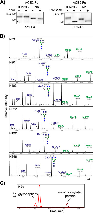
FIGURE 3.

ACE2‐Fc glycosylation. (A) Immunoblot analysis of Endo H and PNGase F digested ACE2‐Fc purified from N. benthamiana and HEK293 cells. (B) MS spectra [M+2H]2+ of glycopeptides from plant‐produced ACE2‐Fc. The assigned N‐glycan structures were labeled according to the ProGlycAn nomenclature (http://www.proglycan.com/). A cartoon illustration highlights the main glycan structure detected for each peptide (http://www.functionalglycomics.org/). (C) Extracted ion chromatogram (EIC) of the most abundant glycopeptides and the non‐glycosylated peptide found on glycosylation site N90 of ACE2‐Fc expressed in N. benthamiana
