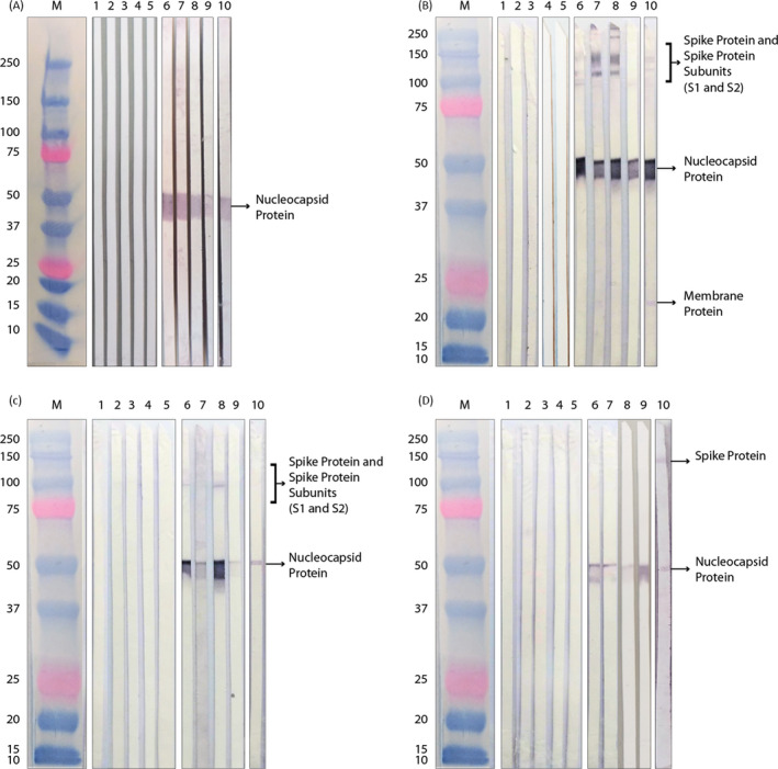
FIGURE 2.

Analysis of structural proteins of SARS‐CoV‐2. Wester blotting of healthy donors (samples 1–5) and patients (samples 6–10) sera, in reducing (A), and non‐reducing condition (B), IgG detection. Presence of IgA (C) and IgM (D) was also detected. The arrows indicate SARS‐CoV‐2 M protein, molecular markers, expressed in kD
