
Figure 3. Comparison of Structural and Functional Parameters of Retina Measured by Swept-Source Optical Coherence Tomography and Multifocal Electroretinography Between Cognitively Normal (CN) Individuals With and Without Amyloid-β Deposition.
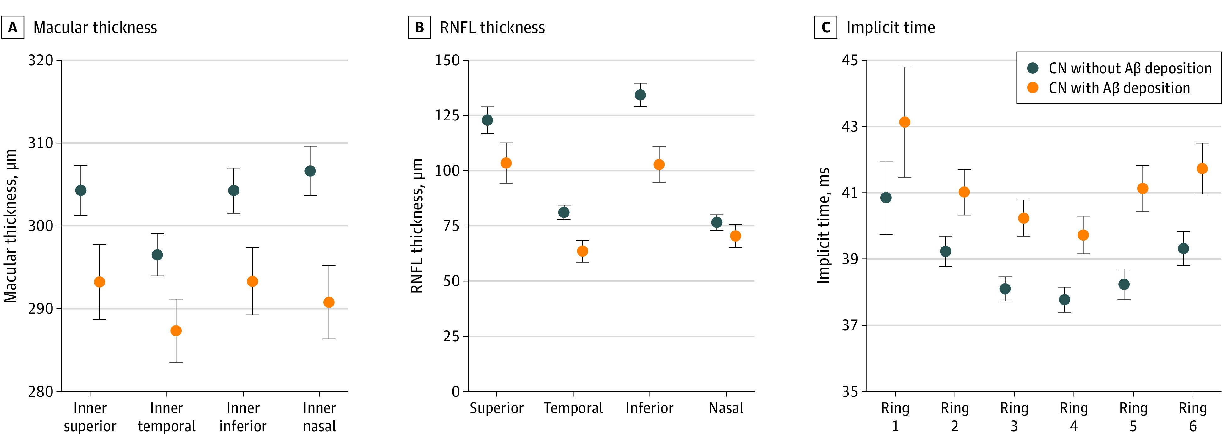
A, Macular thickness of the area in the inner ring of macula. B, Subregional comparison of the thickness of the retinal nerve fiber layer (RNFL) quadrant. C, Implicit time from ring 1 to 6. Dot and error bar indicates adjusted mean and SEM obtained from analysis of covariance after adjusting covariates (age, sex, APOE4, and best-corrected visual acuity).