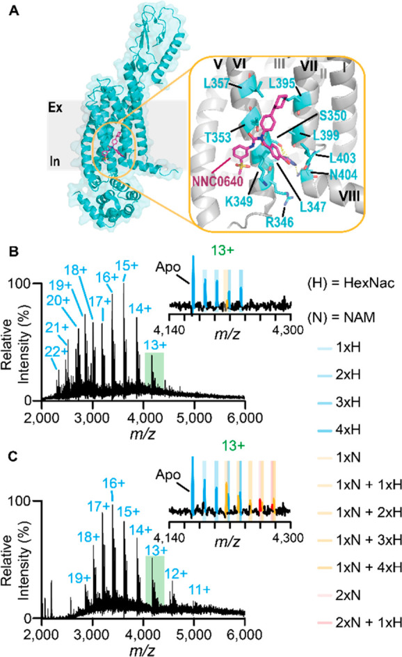
Figure 2.

Preservation of lipophilic drug binding to the glucagon receptor in NaCl-containing electrospray buffers. (A) Structure of the full-length glucagon receptor (PDB 5XEZ) showing the allosteric binding pocket binding NNC0640, a variant of to NNC0666, situated between transmembrane loops VI and VII with interacting residues highlighted in blue. Native mass spectra of GCGR released from G1/CHS micelles in (B) 250 mM NH4OAc, pH 7.5 electrospray buffer or (C) 50 mM NaCl, 5 mM Tris, pH 7.5 electrospray buffers with insets expanding the 13+ charge state. Differences in binding of the NNC066 are more prevalent in (C), highlighted orange and red for one and two NAMs, respectively. Spectra are representative of n ≥ 4 nanoemitters and n = 2 protein preparations.
