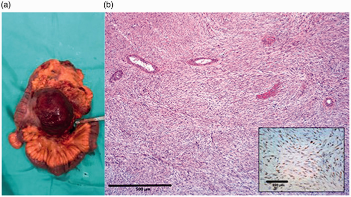
Figure 3.
Macroscopic image of the resected specimen from a 37-year-old female that presented with abdominal pain and a palpable mass in the right hemiabdomen, which shows a well-circumscribed mass that invaded the caecum wall and small bowel mesentery near the terminal ileum without mucosal infiltration (a). Representative photomicrographs of the tumour showing the immunohistochemical staining for beta-catenin. Scale bar 500 µm (inset image, scale bar 250 µm) (b). The colour version of this figure is available at: http://imr.sagepub.com.

