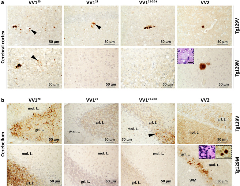
Fig. 3.
PrP immunohistochemistry (IHC). a: Cerebral cortex. 1st row, VV1: PrP granular deposits (arrowheads) often distributed around the rim of vacuoles. VV2: A plaque-like PrP. 2nd row, VV120: PrP deposits co-distributing with SD; arrowhead: granular PrP. VV121 and VV121−20*: negative PrP IHC. VV2: A PrP plaque; inset: H.E. staining of the plaque. b: Cerebellum. 1st row, VV120 and VV121−20*: PrP deposition affecting the granule cell layer (grl. L.); arrowhead, VV121−20*: granular PrP. VV121 and VV2: Negative PrP IHC; mol. L.: molecular layer. 2nd row, VV120: PrP deposition in grl. L. VV121 and VV121−20*: Negative PrP IHC. VV2: Plaque and plaque-like PrP ; dotted and dashed insets: two PrP plaques depicted on H.E. and IHC preparations, respectively. Scale bar insets: 20 µm; Ab: 3F4

