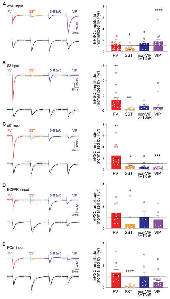
Figure 2. Long-range inputs from diverse brain areas differentially engage different subtypes of GABAergic neurons in the supragranular layers of S1.

Left: example traces of ChR2 photostimulation-evoked EPSCs from PV INs, SST INs, nonVIP-5HT3aR INs, VIP INs, and Pyr from wM1, S2, cS1, ECt/PRh, and POm. Gray traces depict individual sweeps, and solid traces indicate the average of these sweeps. Blue bar indicates ChR2 photostimulation (470 nm, 3 ms). Right: population data showing EPSCs of GABAergic INs normalized to those of simultaneously recorded nearby pyramidal neurons. *p < 0.05, **p < 0.005, and ***p < 0.0005 (Wilcoxon signed-rank test). See also Figures S1–S4.
