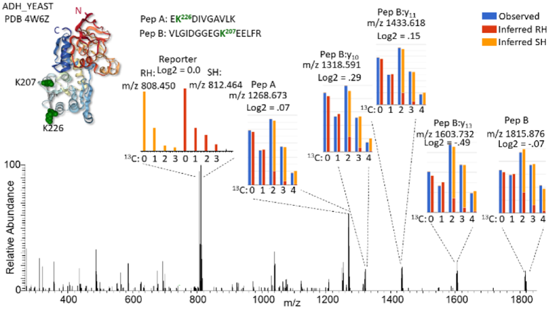
Figure 2.

Example fragmentation spectrum of an iqPIR cross-linked peptide pair. MS2 spectrum of the iqPIR cross-linked peptide pair linking residues 207–226 of ADH1_YEAST a 1:1 mixture of RH/SH cross-linked. The PDB structure 4w6z is shown as a ribbon structure colored red from the N-terminus to blue at the C-terminus with the cross-linked Lys shown as green space filled residues. Insets show expanded views of selected fragment ions, illustrating the isotopic differences which are used for quantification. For fragment ions differing by two 13C, the observed signal is shown in blue while the deconvoluted signal from the RH and SH are shown in red and orange, respectively. The reporter ion signal differs by four 13C, requiring no deconvolution, and follows the red/orange color scheme.
