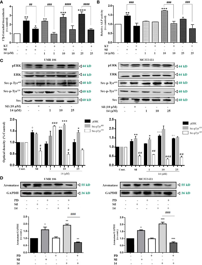
Figure 5.
14 activates PKG/Src/ERK pathway signaling. (A) UMR106 cells seeded in 24-well plates overnight were treated with compounds for 2 h. Subsequently, the cells were supplemented with testosterone (10 nM) for an additional 48 h. 17β-estradiol concentration in the culture medium was quantified with an ELISA (E2) detection kit. (*) p < 0.05, (**) p < 0.01, (***) p < 0.001 and (****) p < 0.0001 compared to the DMSO control; (##) p < 0.01, (###) p < 0.001 and (####) p < 0.0001 compared to KT5823 (10 μM)-treated cells (B) UMR106 cells seeded in 24-well plates overnight were treated with 14 and KT5823 (10 μM) for 2 h. Subsequently, the cells were supplemented with testosterone (10 nM) for an additional 48 h. ALP activity of the cell lysates was quantified with the ALP detection kit. (*) p < 0.05, (**) p < 0.01 and (***) p < 0.001 compared to the DMSO control; (###) p < 0.001 compared to KT-treated cells (C) UMR 106 and MC3T3-E1 cells were treated with different concentrations of 14 and sildenafil (10 μM) for 1 h. The cell lysates were immunoblotted with antibodies against phospho-ERK, ERK, phospho-Src-pTyr418, phospho-Src-pTyr529, and Src. (+) p < 0.05, (++) p < 0.01 and (+++) p < 0.001 compared to the p-ERK control; (*) p < 0.05, (**) p < 0.01, (***) p < 0.001 compared to the Src-pTyr418 control; (#) p < 0.05, (##) p < 0.01 and (###) p < 0.001 compared to the Src-pTyr529 control. (D) UMR 106 and MC3T3-E1 cells were treated with different concentrations of 14 and PD (10 μM) for 1 h. The cell lysates were immunoblotted with antibodies against aromatase. GAPDH was used as the internal control. Cont., DMSO-treated control; E2, 17β-estradiol (10 nM); Sil, sildenafil (10 μM); Rp-pCPT-cGMPS, ERK inhibitor (0.5 mM); KT, PKG inhibitor (KT5823, 10 μM); PD, ERK inhibitor (PD98059, 10 μM). Error bars represent the standard deviation of the measurement. (*) p < 0.05 (**) p < 0.01, (***) p < 0.001 compared to the DMSO control; (###) p < 0.001.

