
FIGURE 3.
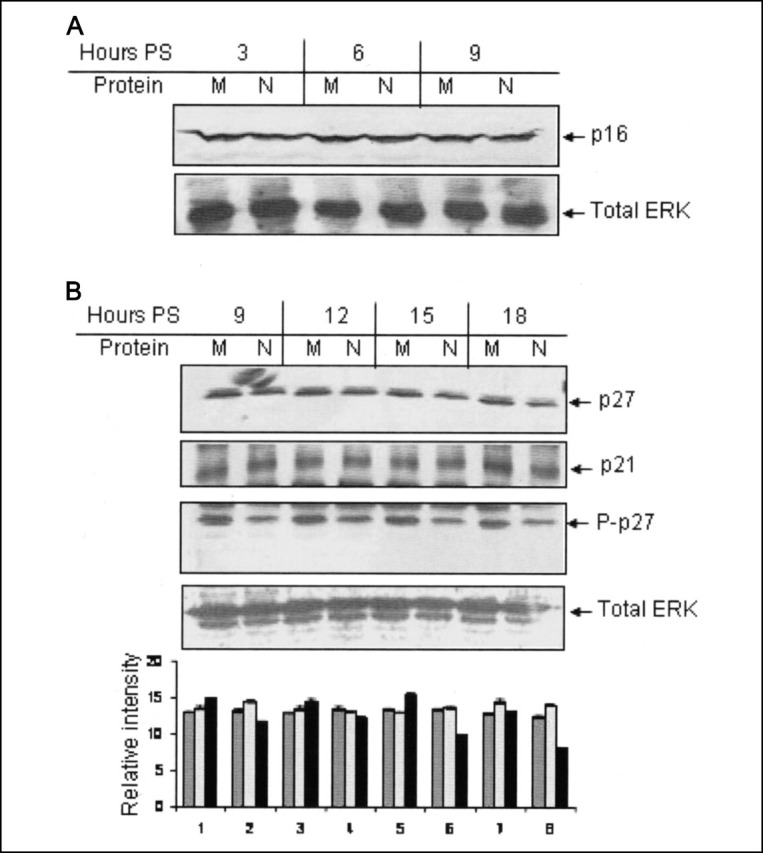
Down-regulation of CDK2 activity is independent of p16, p27, and p21.A, Huh7 cells transfected with pCDN3.1 (M) or pCDNA3.1N (N) plasmid were starved for 34 h, followed by simulation with 10% bovine serum for the indicated time periods. Total cell lysate was immunoblotted with total p16 antibody (first panel). PS, poststimulation. The same blot was stripped and reprobed with total ERK antibody (second panel). B, Huh7 cells transfected with pCDNA3.1 (M) or pCDNA3.1N (N) plasmid were starved for 34 h, followed by simulation with 10% bovine serum for the indicated time periods. Total cell lysate was immunoblotted with total p27 (first panel), total p21 (second panel), and phospho-p27 (Thr187) (third panel) antibody. P-p27 blot was stripped and reprobed with total ERK (fourth panel) antibody. Band intensities were normalized with reference to that of total ERK and graph-plotted. The image is representative of three experiments. In the graph, each set of bars represents the corresponding lane in the gel above; dark gray, light gray, and black bars represent p27, p21, and P-p27 band intensity, respectively.