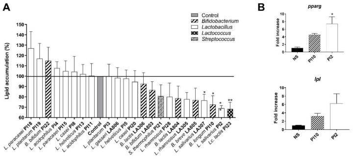
Figure 1.
Effect of selected strains on adipocyte lipid accumulation. (A). Intracellular lipids were quantified after Oil-red-O staining. The dye was eluted from cells with isopropanol and quantified. Results are expressed as a percentage from control adipocytes. * refer to the comparison of probiotic-stimulated 3T3-L1 versus untreated cells (medium control). (B). Relative mRNA expression (RT-qPCR) of pparg and lpl in B. longum PI10- and L. salivarius PI2-treated adipocytes. * refer to the comparison of probiotic-stimulated 3T3-L1 versus untreated cells (medium control), * p < 0.05, ** p < 0.01.

