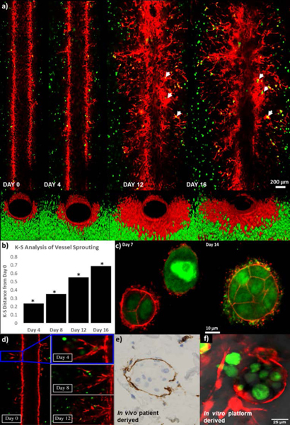
Fig. 6.
Vascular sprouting dynamically observed over a three week period in the MDA-IBC3/TIME in vitro vascularized tumor platforms. (a) Longitudinal cross section images of the vessel show vessel sprouting, branching, as well formation of tumor emboli pointed out by white arrows (top panels), and front view of the vessels (bottom panels). (b) K-S analysis of vessel sprouting revealed a significant increase in sprouting at later time points compared to Day 0. (c) F-actin (red) staining of GFP labeled MDA-IBC3 cells (green) showed formation and growth of tumor emboli. (d) Lumen formation followed over time in one of the vessel sprouts. (e) Vascular nesting phenomenon of IBC tumors in in vivo patient derived histological samples demonstrated by CD31 staining of vascular vessel (brown) surrounding IBC tumor emboli (blue). (f) In vitro recreation of vascular nesting of IBC tumors as shown by the encircling of MDA-IBC3 tumor cells (green) by mKate labeled sprouts (red).

