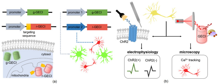
Figure 3.
Multiplexing strategies for GECIs. (a) Multicolor imaging can be done either within compartments of the same cell type or between multiple cell types in vivo and in vitro. This can be done by fusing a targeting sequence to the N-terminus of a red-shifted variant (r-GECI) and introducing this alongside a standard green-variant (g-GECI) to achieve multiplexed Ca2+ tracking in the cytosol and a compartment such as the mitochondria [158]. Another useful application is to utilize cell-type-specific promoters to drive expression of two GECI color variants. This allows simultaneous tracking of Ca2+ levels in multiple cell types in vitro or in vivo, such as driving expression of a g-GECI in astrocytes (promoter 1) and an r-GECI in cortical neurons (promoter 2) of the mouse brain. (b) A cell expressing both light-sensitive ChR2 and a GECI can enable study of Ca2+ dynamics in optically tagged cells [159]. While this optogenetic platform gives an electrophysiological readout of the ChR2-positive neuronal populations in vivo, this can be multiplexed with Ca2+ detection. However, since the commonly used GECIs are also blue-light induced, a red-shifted or NIR variant should be used in order to avoid crosstalk with the optogenetic component of the circuit during laser excitation with fluorescent imaging.

