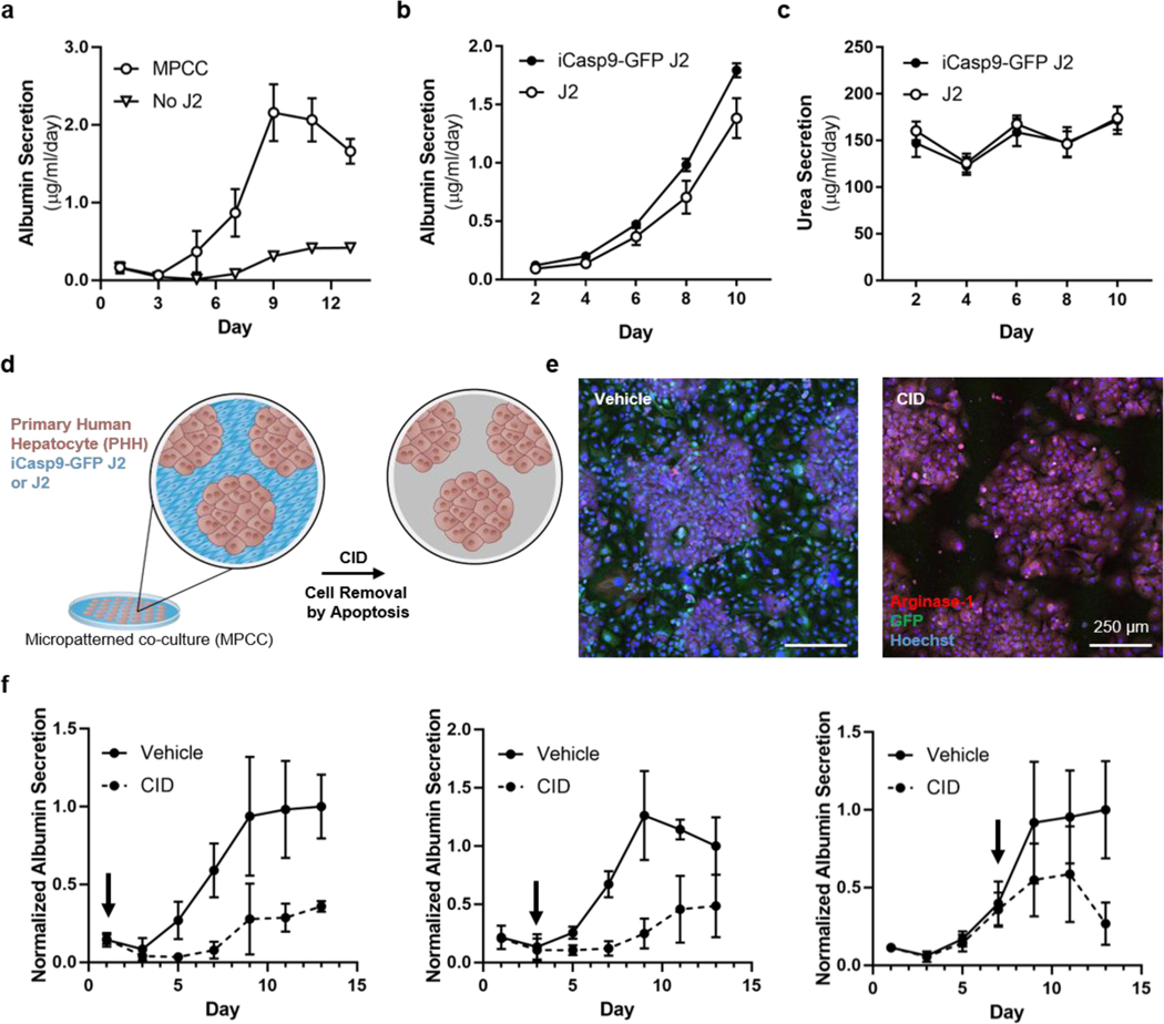
Figure 2. 2D MPCC cultures depend on the sustained presence of stromal cells.
MPCCs or pure hepatocytes were assayed for albumin secretion rate (a, n=3). MPCCs containing wild-type and modified J2s were assayed for albumin secretion rate (b, n=6) and urea secretion rate (c, n=6). MPCCs comprised of primary human hepatocytes (brown) and iCasp9-GFP J2 or wild-type J2 fibroblasts (blue) were treated with CID to remove iCasp9-GFP J2s by apoptosis (d). Vehicle- or CID-treated MPCCs were stained and visualized by immunofluorescence imaging (e, scale bar = 250 μm). MPCCs were treated with CID at day 1, 3, or 7 after initiating co-culture and assayed for albumin secretion rate (f, n = 5, normalized to day 13, arrows indicate CID dose day).

