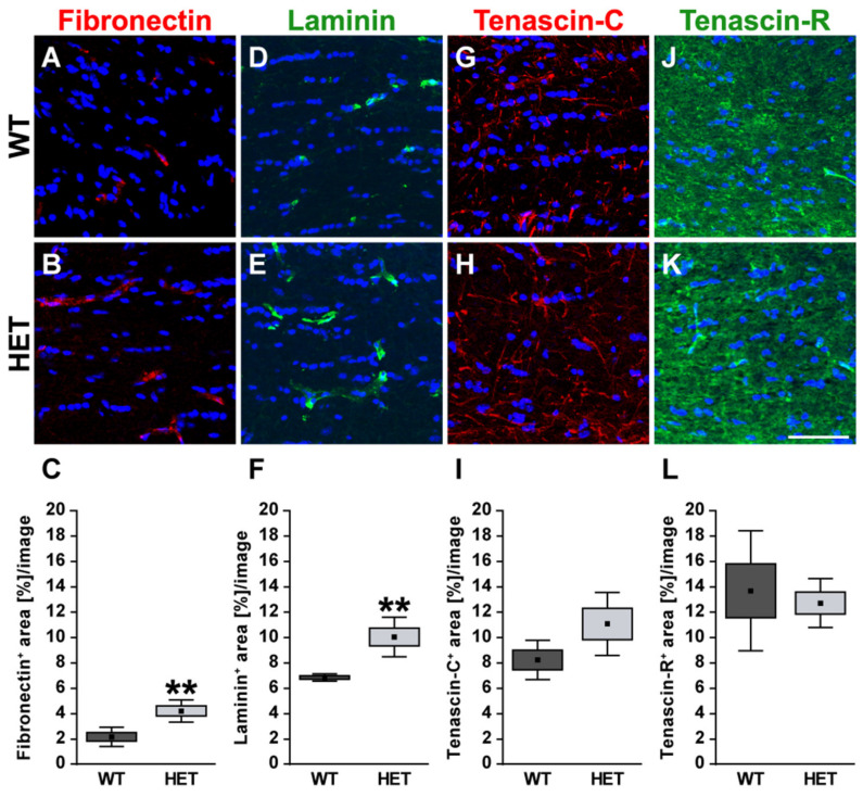
Figure 5.
Enhanced staining signals of blood vessel-associated fibronectin and laminin in the glaucomatous optic nerve of HET mice. Most representative immunohistochemical ECM stainings are shown. Staining of the glycoproteins fibronectin (red; (A–C)) and laminin (green; (D–F)) was significantly increased in the HET optic nerves. Signals were limited to blood vessels. A thread-like-staining pattern was found for tenascin-C (red; (G,H)), while a widely extracellular staining pattern was observed for tenascin-R (green; (J,K)). Both tenascins showed a comparable staining area in the WT and HET group (I,L). Groups were analyzed by Student‘s t-test. Data are shown as mean ± SEM ± SD. ** p < 0.01. n = 5/group. Scale bar = 50 μm.

