Figure 7.
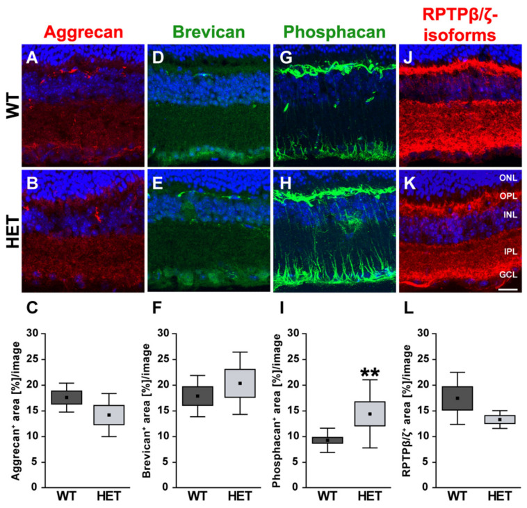
Enhanced phosphacan immunoreactivity in the glaucomatous retina of HET mice. Most representative immunohistochemical ECM stainings are shown. Stainings revealed a prominent signal for aggrecan (red; (A,B)), brevican (green; (D,E)) and all three RPTPβ/ζ isoforms (red; (J,K)) in the plexiform layers and the GCL. The phosphacan signal was restricted to Müller glia fibers (green; (G,H)). Quantification showed a significant upregulation of phosphacan in the HET retina (I). No changes were noted for aggrecan, brevican and all RPTPβ/ζ isoforms (C,F,L). TO-PRO-3 (blue) was used to detect the cell nuclei. Groups were analyzed by Student‘s t-test. Data are shown as mean ± SEM ± SD. ** p < 0.01. n = 5/group. Scale bar = 20 μm. Abbreviations: ONL—outer nuclear layer, OPL—outer plexiform layer, INL—inner nuclear layer, IPL—inner plexiform layer, GCL—ganglion cell layer.

