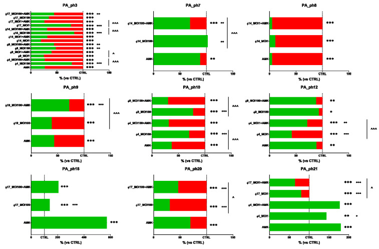
Figure 3.
In vitro phage and amikacin (AMK) activity, alone and in combination, against nine 24-h-old cystic fibrosis Pseudomonas aeruginosa (PA) biofilms. Biofilm dispersion evaluated by crystal violet stain. The results are shown as percentages in dispersed PA biofilms (highlighted in red) compared with unexposed PA control samples (CTRL) in trypticase soy broth (TSB); the dotted line indicates 100% residual biofilm after a challenge with TSB; significant levels * p < 0.05, ** p < 0.01, *** p < 0.001 each treatment vs. CTRL; ° p < 0.05, °° p < 0.01, °°° p < 0.001 each treatment vs. AMK, and ^ p < 0.05, ^^^ p < 0.001 each treatment vs. phage-AMK analyzed by the χ2 test.

