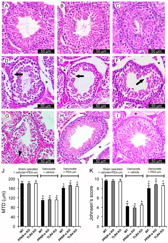
Figure 1.
Histological organization of the testes with Hematoxylin-Eosin stain. (A–C): The images and the means ± SE of only one of the control groups (sham operated + vehicle and sham operated + N-Palmitoylethanolamide (PEA)) are provided, as to not overcomplicate the figure. A normal structure of both the tubular and the extratubular compartments is evident. (D–F): Wild-type (WT), peroxisome proliferator-activated receptor-α knockout (PPAR-α KO), toll-like receptor 4 knockout (TLR4 KO), varicocele operated mice treated with vehicle, respectively. Tubules with discontinuous epithelium (arrow) and marked extratubular edema (*) are demonstrated. (G): WT varicocele operated mice treated with ultra-micronized PEA (PEA-um). Significantly larger tubules with elongated spermatids can be observed. Extratubular edema (*) is reduced and blood vessels (arrowhead) show mild dilation. (H): PPAR-α KO varicocele operated mice treated with PEA-um. The germinal epithelium shows a close to normal organization, even if an evident edema (*) is present in the extratubular compartment. (I): TLR4 KO varicocele operated mice treated with PEA-um. The germinal epithelium has a normal structure with many spermatids and mature spermatozoa. A mild edema (*) is present in the extratubular compartment. (J): Quantitative evaluation of the mean tubular diameter (MTD) in the different groups of mice. (K): Johnsen’s score in the different groups of mice. * p < 0.05 vs. sham operated groups; ° p < 0.05 vs. varicocele plus vehicle groups. (Scale bar: 50 µm).

