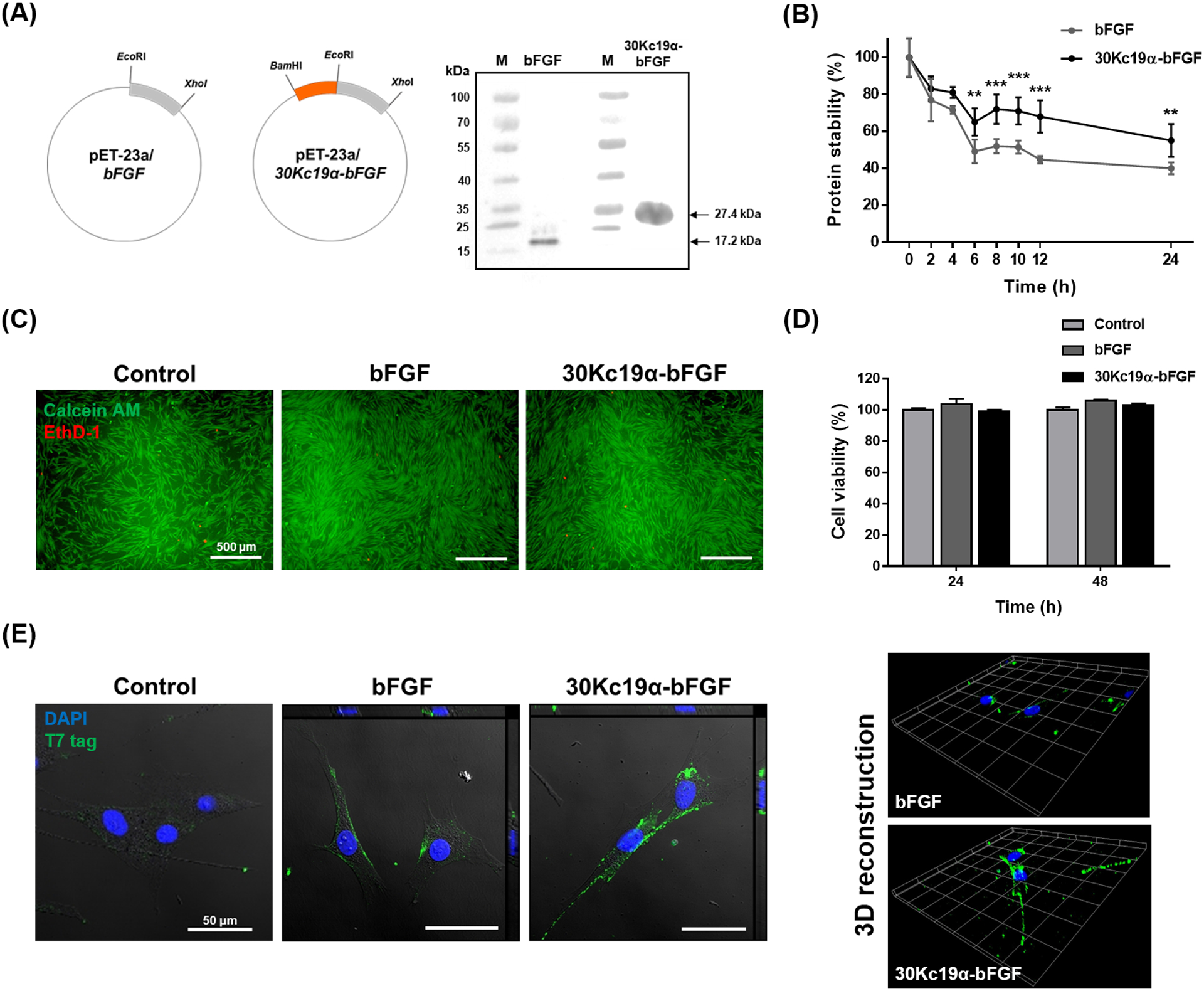
Figure 1. The Fusion of 30Kc19α to bFGF and Its Effects on Stability and Cell-Penetration of bFGF.

(A) Plasmid construction of bFGF and 30Kc19α-bFGF and western blot analysis of recombinant proteins. Anti-T7 tag antibody was used as a primary antibody. (B) Protein stability assay using the bFGF ELISA kit. Proteins at 10 nM were incubated at 37°C from 0 to 24 hours. The relative protein stability was obtained by normalizing protein stability at each time point to protein stability at t = 0 (**p < 0.01, ***p < 0.001 compared with bFGF group). (C) Cytotoxicity assay using LIVE/DEAD kit and (D) CCK-8 kit. Human dermal fibroblasts (HDFs) were treated with 4 μM of the proteins for 24 and 48 hours. Live and dead cells from 48 hour treated samples were stained with green and red fluorescence respectively (Scale bar=500 μm). CCK-8 assay was done for both 24 and 48 hour treated samples. Data are expressed as relative cell viability normalized to the control group (n=4). (E) Confocal image with orthogonal projection (left) and 3D recostruction of z-stack images (right) of bFGF- and 30Kc19α-bFGF-treated HDFs. HDFs were treated with 4 μM of the proteins for 1 hour. The proteins were tagged with Alexa Fluro® 488, and the nucleus with DAPI (Scale bar = 50 μm).
