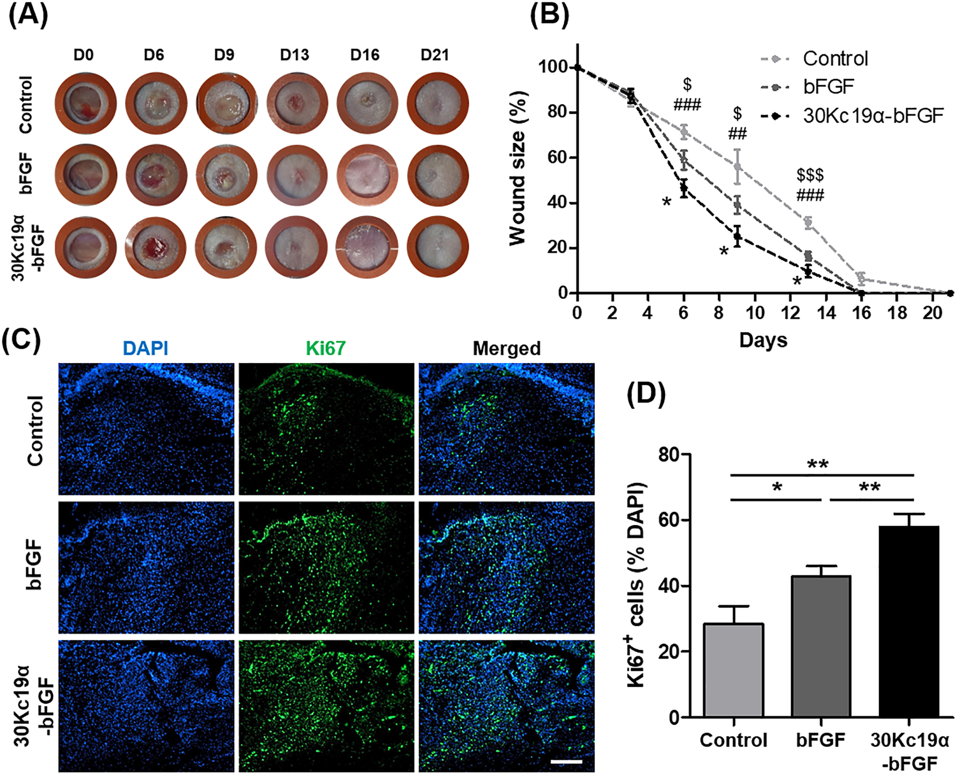
Figure 6. In Vivo Wound Healing Application of 30Kc19α-bFGF.

(A) Photographs of the wound up to 21 days. (B) The wound size-reduction profile was calculated based on the photographs. 30Kc19α-bFGF significantly promoted wound healing compared to bFGF (Statistical significance: * represents 30Kc19α-bFGF to bFGF; # represents 30Kc19α-bFGF to control; and $ represents bFGF to control. *,$p<0.05, ##p<0.01, and ###,$$$p<0.001). (C and D) Proliferative cells in the wound bed were estimated by immunofluorescence staining of Ki67 (green) on day 6, and quantitatively analyzed based on the fluorescent signals. (Scale bar=200 μm) (n=5–8) (*p<0.05 and **p<0.01,).
