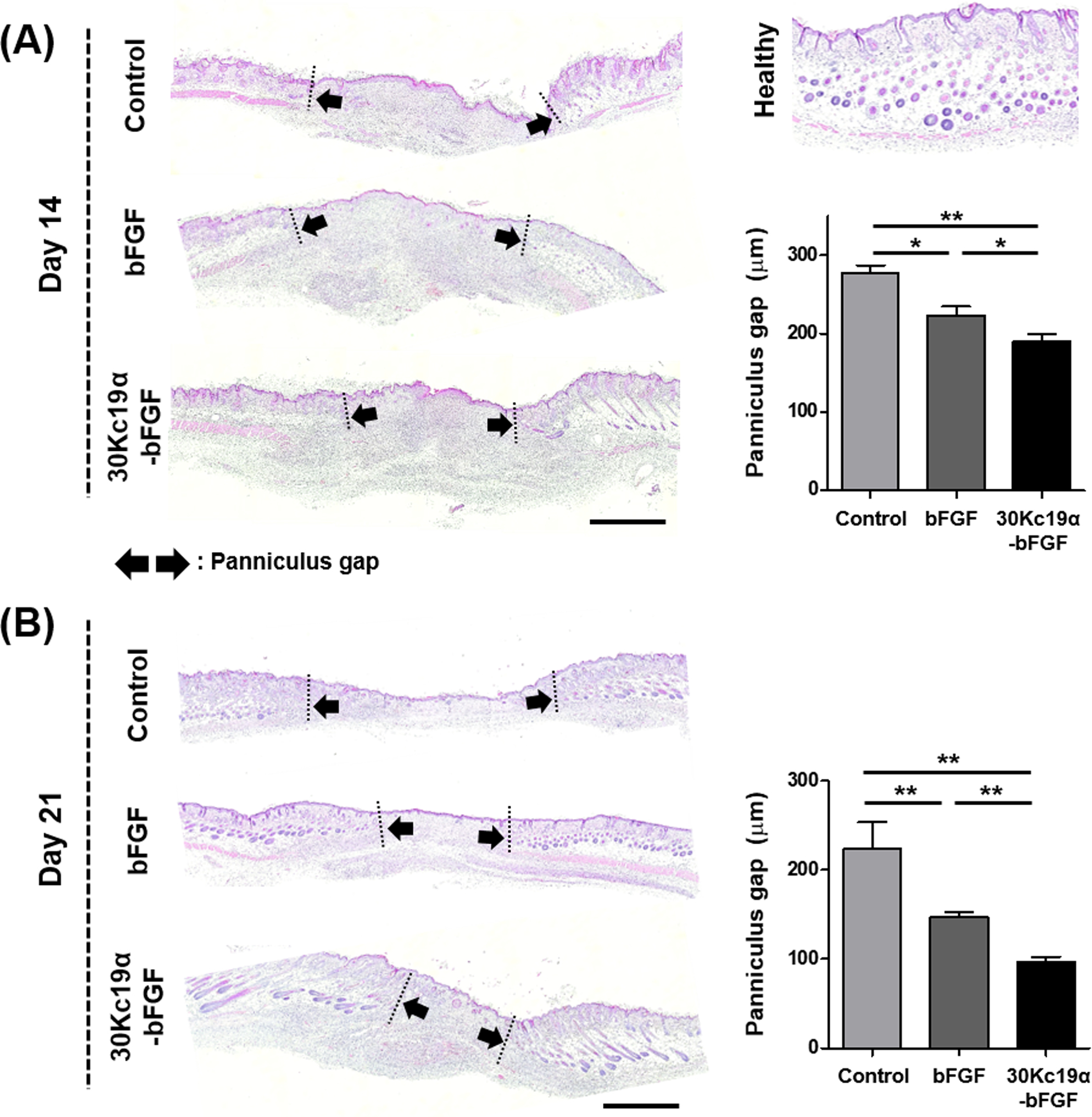
Figure 7. Histological and Qualitative Analysis Based on Hematoxylin and Eosin (H&E) Stain.

Panniculus gap was quantified on day 14 (A) and day 21 (B). It was revealed that 30Kc19α-bFGF accelerated wound regeneration via tissue granulation process. Although the panniculus gap of all groups continuously decreased, it was notably reduced with 30Kc19α-bFGF, whose tissue was being recovered at the fastest rate with a structure similar to healthy tissue (Scale bar=1 mm) (n=3–4) (*p<0.05 and **p<0.01).
