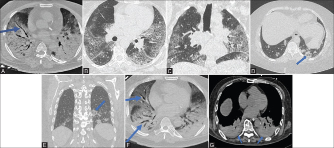
Figure 3 (A-G).
(A): CT chest (axial) shows markedly enlarged vessel (solid blue arrow) against a backdrop of consolidations and GGO in a COVID-19 patient. Acute respiratory distress syndrome with dense consolidations is seen on both sides. (B and C) Axial and coronal images show bilateral interstitial septal thickening with background ground glass giving the appearance of “crazy paving.” (D and E): axial and coronal images from CT chest of a patient showing circular consolidation with GGO within in the posterior, peripheral location, abutting the pleura in the left lower lobe (solid blue arrow). (F) In the same patient of image 3A, this CT chest (axial) shows bilateral bronchiectasis (solid blue arrows) against a backdrop of bilateral consolidations and ground glass. Acute respiratory distress syndrome with dense consolidations is seen on both sides. (G) CT chest (axial) shows bilateral pleural effusion at the lung bases (solid blue arrows). Acute respiratory distress syndrome with dense consolidations is seen on both sides

