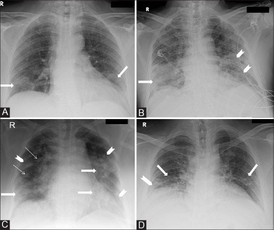
Figure 1 (A-D).

CXR AP views of four different COVID-19 patients at presentation demonstrating various specific findings. (A) Subtle GGOs (arrows) are seen in bilateral lower zones. (B) Consolidation (arrow) is seen in the right lower zone and GGOs are seen in left lower zone (arrow heads). (C) Consolidations are seen in the bilateral lower zones and left mid zone (thick arrows); peripheral GGOs (arrow heads) are seen bilaterally and nodules (thin arrows) are seen in the right mid zone. (D) Reticular opacities are seen in bilateral lower zones (arrows) along with small GGOs in the right lower zone (arrow head)
