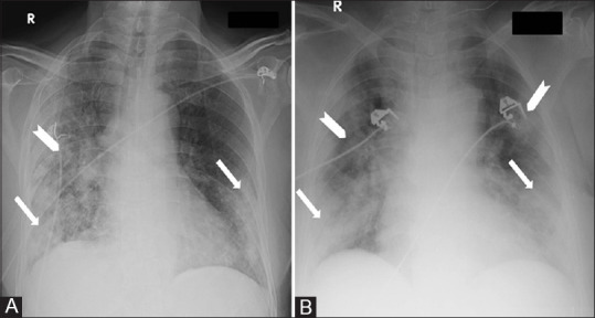
Figure 3 (A and B).

CXR AP views of two different COVID-19 patients demonstrating asymmetrical and symmetrical abnormalities. (A) 72 Y/M presented with fever and malaise since 10 days. CXR shows bilateral lung parenchymal abnormalities (right more than left) with areas of bilateral lower zone consolidations (arrows) mixed with right middle zone GGOs (arrow head). (B) 64 Y/F with history of diabetes mellitus presented with fever and dry cough since six days. CXR shows bilateral symmetrical lung parenchymal abnormalities with areas of consolidations (arrows) mixed with GGOs (arrow head)
