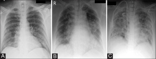
Figure 4 (A-C).

CXR AP views of three different COVID-19 patients demonstrating radiographic grading of severity of disease. (A) Mild grade: small areas of GGOs occupying bilateral lower zones and the abnormal white area is less than the normal black area. (B) Moderate grade: GGOs seen in bilateral peripheral and central lung parenchyma and the areas of white and black are equal. (C) Severe grade: GGOS seen diffusely infiltrating the lung parenchyma and the white area is more than the black area
