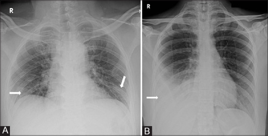
Figure 5 (A and B).

(A) CXR AP view of a 30 Y/M asymptomatic COVID-19 patient showing small GGOs in bilateral lower zones (arrows). (B) CXR AP view of a 32 Y/F presenting with fever and cough since fivedays and history of contact with COVID-19 patient shows pleural effusion on right side without any other specific lung parenchymal abnormality
