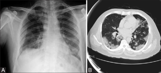
Figure 3 (A and B).

(A) CXR of 44-year-old patient shows bilateral peripheral air space opacities predominantly bilateral lower zones - Typical COVID-19 (Xray severity score of 2/8). (B) Axial HRCT Chest shows a typical pattern for COVID-19 with CT severity score of 11/25
