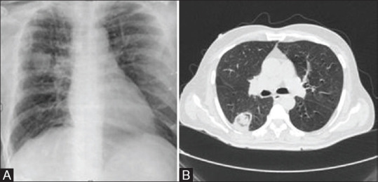
Figure 6 (A and B).

(A) CXRshows rounded mass-like consolidation in right mid lung zone—proved as fungal pneumonia later on HRCT chest- NON-COVID-19 pattern.(B) Axial HRCT chest shows thin-wall cavity within apical segment of the right lower lobe with soft tissue density lesion suggestive of fungal etiology
