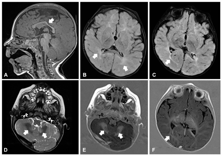
Figure 2.
Brain magnetic resonance imaging in patient 1 at age 10 months. Corpus callosum is thin and mildly dysplastic at the level of isthmus (arrow in (A), sagittal view, T1-weighted image). The deep and periventricular white matter appears hyperintense compared to the grey matter (arrows in (B), axial view, FLAIR image), a similar abnormal high signal also involves the optic radiations (arrow in (C), axial view, FLAIR image). Finally, mild hyperintensity of the cerebellar dentate nuclei is visible (arrows in (D), axial view, T2 weighted image). T1-IR images confirm the involvement of dentate nuclei (arrows in (E), axial view) and optic radiations (arrow in (F), axial view).

