Figure 1.
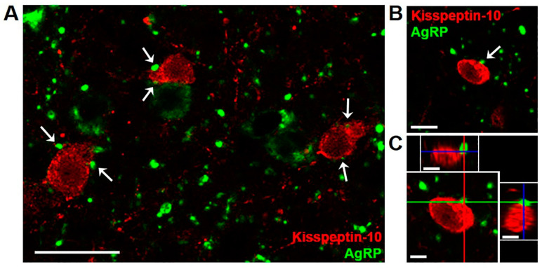
Confocal images (1.0 µm optical section; 40 × magnification) showing dual-label immunofluorescence for AgRP (green) and kisspeptin (red) in the arcuate nucleus of a castrated male sheep (wether). White arrows (A,B) indicate examples of AgRP-immunoreactive (ir) terminals in apposition to arcuate kisspeptin neurons, with AgRP-ir cell bodies and fibers seen in the vicinity. Orthogonal views (C) confirm close contact of an AgRP-labeled bouton to a kisspeptin cell body. Red, green, and blue lines in (C) indicate X, Y, and Z planes, respectively. Scale bars = 25 µm (A), 10 µm (B,C).

