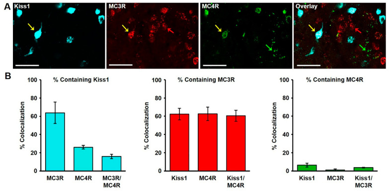
Figure 2.
(A) Confocal image (1.0 µm optical section; 20 × magnification) of kisspeptin, MC3R, and MC4R mRNA-expressing cells in the arcuate nucleus (ARC) of a castrated male sheep. Yellow arrows indicate a Kiss1 cell expressing both MC3R and MC4R. Red and green arrows show MC3R and MC4R-expressing cells, respectively. Scale bar, 50 µm. (B) Mean (± SEM) percentage of kisspeptin (left), MC3R (middle), and MC4R (right) cells containing Kiss1, MC3R, and/or MC4R in the middle ARC.

