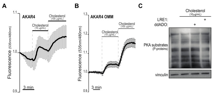
Figure 3.
Cholesterol supplementation induces rapid PKA activity predominantly in proximity of sub-cellular organelles. (A,B), Pancreatic acinar cells were isolated from wild-type animals, infected with adenoviral vectors encoding for the PKA sensors AKAR4 (that senses cytoplasmic PKA activity, panel (A) and AKAR4-OMM (that targets PKA activity localized on the surface of intracellular organelles, panel (B). After 36 h, cells were embedded in a synthetic matrix and imaged. Fluorescence intensities were recorded for several minutes, before and after the addition of indicated amounts of cholesterol. The graphs show FRET ratios over time. Scale bars, 3 min. In (C), primary acinar cells were added cholesterol or vehicle and immediately treated with indicated inhibitors (30 µM both) for 15 h. Whole-cell lysates were blotted to detect phosphorylated PKA substrates.

