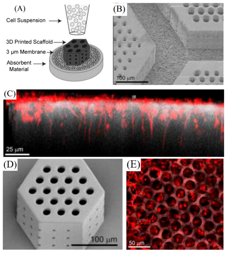
Figure 7.
(A) Schematic of the retinal progenitor cell loading strategy. (B) Large photoreceptor cell porous membrane adhered to the membrane after processing. (C) Side view of retinal neurons (marked in red) settled in and aligned with 25 μm vertical pores. (D) Representative scanning electron microscopy (SEM) image of a small retinal progenitor cell scaffold used to determine design-to-structure fidelity. (E) Sequential top-down images of retinal neurons (marked in red) on the surface of photoreceptor scaffolds and nestled in 25 μm pores. Reproduced with permission from [57], Elsevier, 2017.

