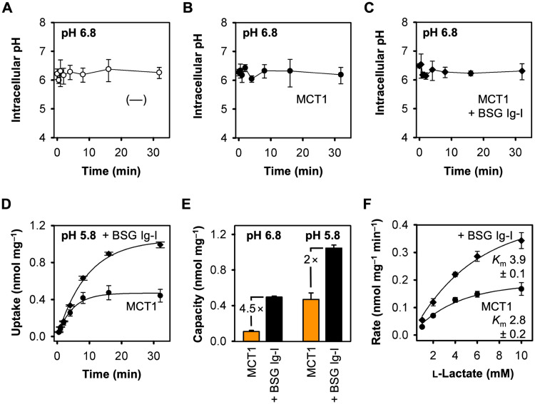
Fig 3. Effect of the transmembrane proton gradient and altered substrate concentrations on transport via MCT1 in the presence and absence of the basigin Ig-I domain.
(A-C) Effect of adding 1 mM l-lactate at an external buffer pH of 6.8 to yeast cells without monocarboxylate transporters (A, 〇), or expressing MCT1 (B, ●) or MCT1 fused with BSG Ig-I (C, ◆). (D) Uptake of 14C-labeled l-lactate via MCT1 alone (●) and fused wit BSG Ig-I (◆) at an external pH 5.8 and a 1 mM inward l-lactate gradient. (E) Uptake capacities of cells expressing MCT1 alone (orange) and fused with BSG Ig-I (black) at pH 6.8 (based on data from Figs 1D and 2C) and pH 5.8 (data from 3D). (F) Michaelis-Menten kinetics for MCT1 alone and fused with BSG Ig-I. Km values were determined at pH 6.8 and are given in mM. In all cases, the background of non-expressing cells was subtracted; error bars indicate ± S.E.M. of three biological replicates.

