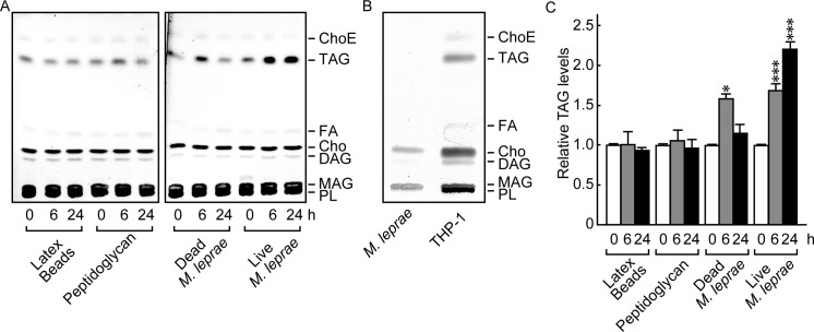
Fig 1. M. leprae infection enhanced TAG accumulation in THP-1 cells.
(A) THP-1 cells (3 × 10⁶) were cultured in 6-well plates with live M. leprae (MOI: 20), heat killed (80°C for 30 min) M. leprae, 2 μg/ml peptidoglycan, or latex beads for the indicated period of time. Total lipids were extracted and analyzed by HPTLC. (B) Total lipids extracted from M. leprae and THP-1 cells were analyzed by TLC. (C) The amount of TAG as measured by densitometry and expressed relative to levels at 0 h. Values represent the mean ± S.D. from three independent experiments. Significance was determined by a one-way ANOVA followed by a Dunnett test. One and three asterisks indicate p<0.05 and p<0.001, respectively. ChoE: Cholesterol ester; TAG: Triacylglycerol; FA: Fatty acid; Cho: Cholesterol; DAG: Diacylglycerol; MAG: Monoacylglycerol; PL: Phospholipid.

