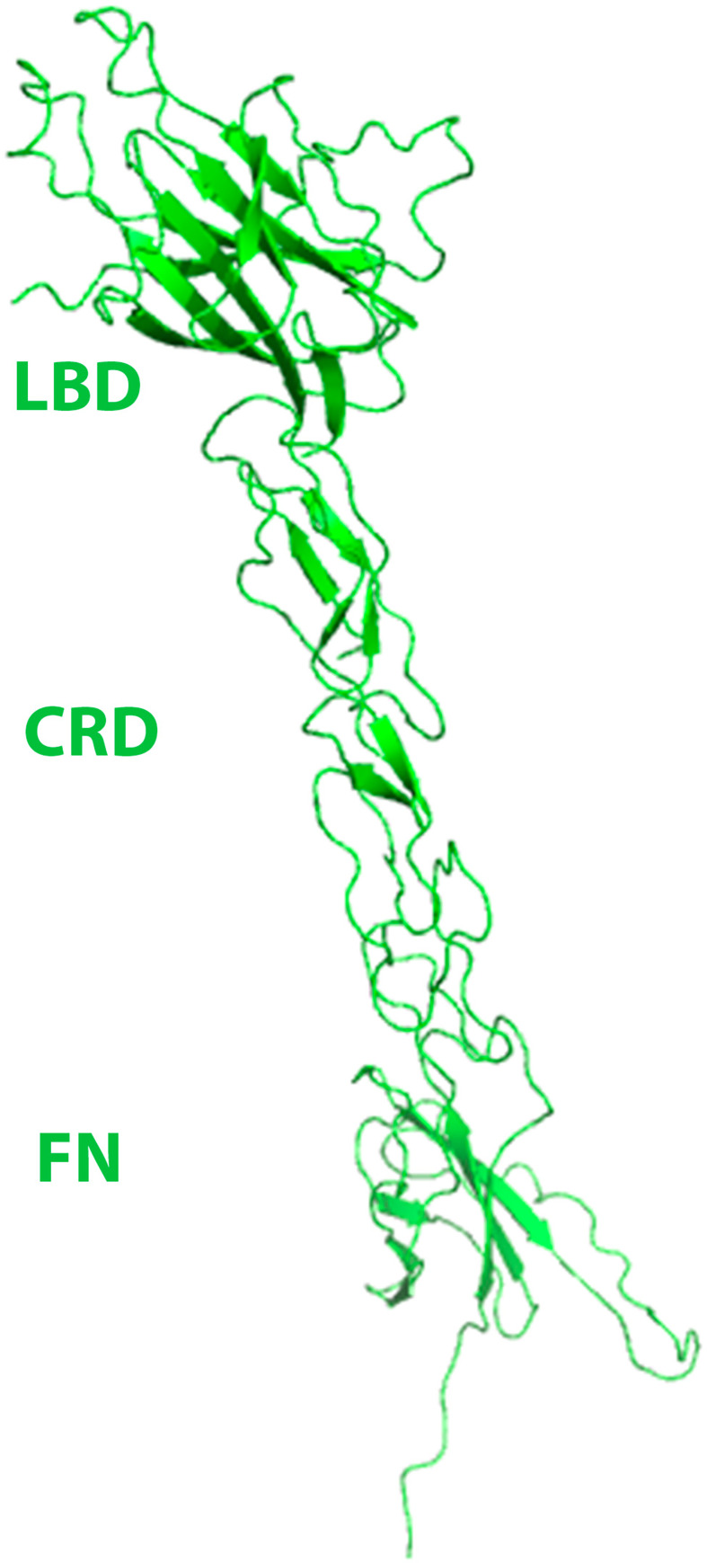
Fig 4. EphB6-ECD structure showing the ligand binding (LBD), cysteine rich (CRD), and fibronectin (FN) domains.
The ectodomain has a rigid, rod-like conformation with limited flexibility between the subdomains. The LBD has a jelly-roll folding topology, while the CRD includes β-strands arranged as a β-sandwich, with several disulfide bonds stabilizing the structure. The N-terminal FN adopts a typical immunoglobulin-like fold.

