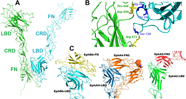
Fig 6.
(A) Head-to-tail interactions in EphB6-ECD. Similar to the previously published A-class ectodomain structures, the LBD (cyan) and FN (green) domains of neighboring EphB6 molecules form an interface in the unliganded receptors. (B) The zoom-in shows the interacting amino acid residues. (C) Comparison of the head-to-tail interactions in EphB6-, A4- and A2-ECDs. (Left) EphB6-LBD (cyan) bound to EphB6-FN of a neighboring molecule (yellow) in the crystals of the unliganded EphB6-ECD; (Center) EphA4-LBD (blue) bound to the EphA4-FN of a neighboring molecule (orange) in the crystals of unliganded EphA4-ECD; (Right) EphA2-LBD (green) bound to the EphA2-FN of a neighboring molecule (red) in the crystals of unliganded EphA2-ECD.

