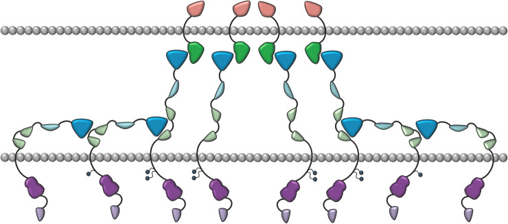
Fig 7. Schematic representation of the head to tail Eph-ECD interactions.
The Eph receptors are in blue/green/purple and are interacting with the ephrins in green/orange. The LBDs of the ephrin-free Ephs (in blue) and the FN3 regions (in light green) of their neighbors are interacting. Intracellular, juxtamembrane Tyrosines are shown as small circles. They get fully phosphorylated only when biologically active heterotetramers and higher-order Eph/ephrin assemblies form after the initial ligand binding events. The cell membrane of the two interacting cells are in grey.

