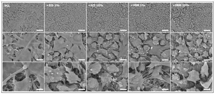
Figure 7.
Representative scanning electron microscopy images of human conjunctival epithelial cells cultured on electrospun scaffolds fabricated from poly(ε-caprolactone) (PCL) and PCL with addition of 1% or 10% decellularised tissue powder (small intestinal submucosa (SIS) and urinary bladder matrix (UBM)). Images: top row = ×1000 (scale = 50 μm), middle row = ×5000 (scale = 10 μm), bottom row = ×10,000 (scale = 5 μm); arrows indicate presence of microvilli.

