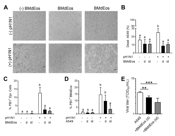
Figure 4.
Eosinophils protect the epithelial cell barrier during influenza virus infection. (A) Representative images of A549 cell monolayers in each culture condition. (B) Percentage of dead A549 cells in the culture conditions. (C) Influenza internal protein (PB1) expressing A549 cells and (D) bone marrow-derived eosinophils in each culture condition. (E) Influenza A virus (pH1N1) titer in the supernatants. Experiments independently repeated three times for rigor and reproducibility. Data are represented as the mean and standard deviation of n = 5–6 samples analyzed by two-way ANOVA with Sidak’s multiple comparisons test. Differences are significant (p < 0.05) when letters above bars are dissimilar. ** p < 0.01 and *** p < 0.001. BMdEos—bone marrow-derived eosinophils; d/D—direct contact; id/ID—indirect contact.

