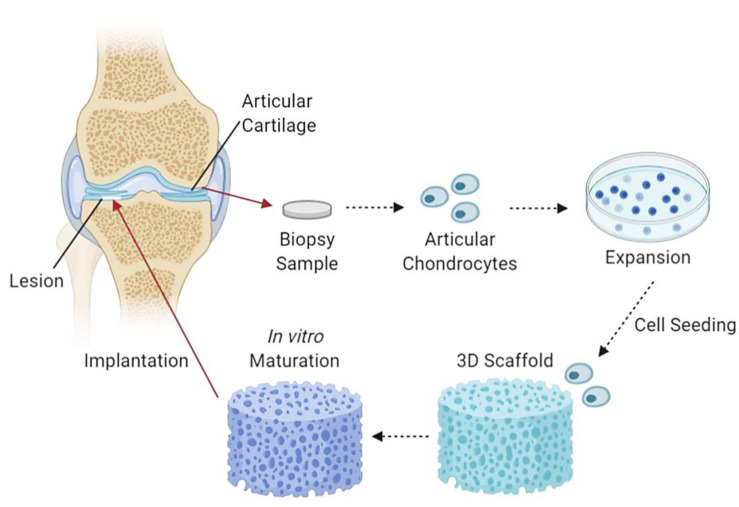
Figure 4.
An illustrative flow diagram of cartilage tissue engineering. It begins with taking a biopsy sample of articular cartilage containing articular chondrocytes, which are then cultured, seeded and incorporated into a 3D scaffold, left to mature partially in in vitro conditions and followed by scaffold implantation into chondral lesions [116]. Created with Biorender.com (Accessed date: 10 February 2021).

